What is medical image management?
A collective term used to describe the management of all types of medical images within the clinic or healthcare organization.
Modern medical image management is done with computers and software. A medical image management system should work enterprise wide – all disciplines of healthcare.
Modern medical image management provide, at least, these benefits for your clinic or organization.
- Recording of images and video from imaging equipment (endoscopes, microscopes, cameras etc.)
- Collaboration around live and recorded images with purpose made functions and features
- Workflow optimization that makes the management streamlined and hassle free
- Automation of common tasks to help save time and resources as well as create high data quality within the workflow

Who needs medical image management?
All healthcare establishments using image producing equipment need a solution to handle their images in order to make good use of them, transform them into an asset for the organization and provide modern, safe and secure healthcare. Hence – most healthcare establishments.
Through a modern, connected medical image management solution you get
- Great workflow structure and effectiveness
- Security and integrity for the patient and his data
- Superb documentation productivity
- Top notch documentation quality and accuracy
- Image based collaboration pathways within the organization
- Modern and better healthcare in other words!
Read more
When we talk about medical image management we refer to working with medical images. Our customers, however, often ask about medical image documentation. Most of the time , what they mean is medical image management. Please take a moment and reflect on the differences and consider what you actually refer to!
Medical image management is a collective term describing the handling of all types of images within the healthcare environment.
Today’s healthcare systems are filled with image producing equipment. Endoscopic units, microscopes, camera units in the OR-lighting or perhaps on the head of the surgeon, surgical robots and so on. Most of these units are used for visualization and image-based guidance – as an extended eye of the physician.
Despite the high presence of image producing equipment, still very few clinics have well established routines for documenting (recording) images and video to create a complete and objective record of the examination or treatment. This is not due to lack of will or interest but rather due to lack of a structured or automated way of doing so. This is where image management solutions make a difference!
Distributed Medical offers a solution for collecting, storing, editing, and sharing medical images and video in an automated, workflow optimized, secure and clinically relevant way.
We call this solution VidiView Enterprise Imaging. It is based on the network infrastructure already in place – in combination with our tailor-made software. This combination makes up a powerful tool for medical image management. The ability to distribute images and video around – instantly – is the one feature that makes it future proof, scalable and a solution you can grow and evolve with. The software is tailor made for and with medical professionals to make it as suitable as possible for the job!
Also, of great importance, is the fact that medical image management is our core business. In fact, our only business. In fact, we find that smooth image and information distribution is so important and relevant that we decided to include it in the name of the company…
That is how committed we are to help improve your medical image management process!
Common VidiView use cases
The VidiView system is not limited or made for a specific, clinical purpose but rather ‘all purposes’ of image management and image capture in the clinical space. It is very customizable and will fit most scenarios, with the exception of the radiology space which it usually rather integrates with.
Below you find five common deployment scenarios which are all proven in combat and easy to deploy in most similar environments.
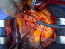
Thoracic surgery
Often in cases with complex, invasive surgery the need for standardized documentation is not always obvious. If you could have a solution that automagically documented unexpected finds or tricky situations that would be great. Most surgeries require none or little documentation though.
An intuitive visualization solution – allowing you to move images around to different screens available in the room would be great though!
All of this, and much more, you can have with VidiView!
- With its SmartRecording-tool set you may start documentation up to 1 hour after something happened. You can record the past!
- With the AVR-tool set you can control various brands of visualization infrastructure (monitors and switches) and by our intuitive interface easily move images around to screens inside the room.
- With VidiViews multi-channel capacity you may connect to 8 different image sources into the system. Valuable in complex ORs like a heart surgery suite.
All of this functionality is packed into one monitor-size unit and presented through a beautiful and easy-to-learn user interface made for healthcare personal.
But do not take our word for it – request a demo and experience this yourself!
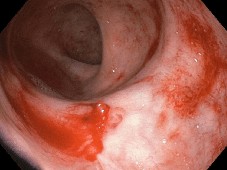
Colonoscopy
Within the gastro area and in colonoscopy especially, images have always been very central. Today the need for video is equally common as the demand for collaborative healthcare is growing all the time.
Since the entire examination procedure is camera driven and since the imaging equipment on to market ranges from standard video all the way up to fantastic 4K UHD video sensors the quality of the images and videos documented during these procedures is very high.
- Modern image management does not only give you easy-to-use documentation tools. It opens possibilities for collaboration as well. Ask a colleague to join your examination for a second opinion!
- Many colonoscopy examinations are carries out in standardized ways. Make use of smart and intuitive anatomy mapping to ensure you have images from all vital parts of the anatomy!
- Use clip recording! -10 seconds to +10 seconds from the press of a button give you live images instead of the static, still images you are used to. Easy to create! Easy to review! Easy to auto archive!
- A great way to do work with images and save valuable resources at the same time in screening programs might be to produce standardized image based documentation during your screening examinations. Examination with uncertain outcome can later be reviewed by a senior physician for possible recalls.
Running a modern gastro unit with a modern image management system is the best way to ensure high quality outcome, rigid documentation routines and best in class patient safety!
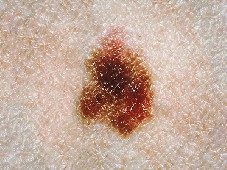
Dermatology
Within dermatology the image has always been very central. The image of the eye as well as the image kept for records. Still image photography has been used for a long time within the field and is often done with a special dermascopic lens, a light source, and a rather conventional camera unit such as a Canon EOS-body.
Till now. With our mobile image capture solution, based on the most sophisticated piece of technology you carry around – you smartphone, you have a very powerful imaging unit! For best performance and workflow, a dermascopic addon should be mounted over the camera lens of the phone. Various models of such addons exists on the market and have been tested with the solution.
- Enjoy a integrated worklist for secure patient data in your exams
- Experience the benefit of the connected healthcare – your photos will automatically be in your computer for review before you sit down! No more manual import and patient data input.
- Map your photos with anatomic information as they are taken
- Map your photos with diagnostic information as they are taken
All together you will make your exams more accurate, secure and effective. Save up to 75% of the time it takes to document your dermatology exams today!
But do not take our word for it – request a demo and experience this yourself!
Dermatology
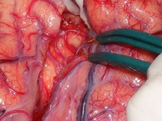
Neurosurgery
Often during neurosurgery imaging equipment of various kinds are crucial to the entire procedure. Often documentation is thought of as equally important. This is usually due to lack of video documentation capabilities and very long procedures.
With our image management solution this is not an issue. You may connect as many image sources as you like (up to 8 individual sources) and you may record the entire procedure – regardless of length. Really!
Aside from documentation you may want to visualize the procedure for staff in the room, not being able to work as close as yourself with the patient. Such visualization might provide important information to the anaesthesiologist or the supply nurse – staying on par with your work. From the same, intuitive touch panel interface you control visualization – on as many screens around the room as you like.
This kind of documentation and visualization will leverage your performance in the OR. At the same time it will as provide an objective, secure and high quality track record of your clinics work with minimal extra work and effort!
But do not take our word for it – request a demo and experience this yourself!
Neurosurgery
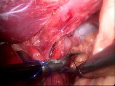
Abdominal surgery
Today a large part of the abdominal surgeries are laparoscopic procedures. Effectively the laparoscope is an extended eye of the surgeon. A true “camera-inside-the-body” procedure where documentation and telemedic possibilities are apparent.
Despite these favourable conditions, far from all laparoscopic suites are equipped with a modern image management solution. Some are. Some are still using thermal printers for documentation and have no tools at all for collaboration with colleagues. With todays very accurate and high performing laparoscopic equipment providing ultra high definition video the ability to document and collaborate with great accuracy is better than ever.
- Modern image management does not only give you easyto-use documentation tools. It opens possibilities for collaboration as well. Ask a colleague to join your examination for a second opinion!
- Document the entire procedure for later review in case of problems during patient recovery.
- Document with video and audio for reconstruction purposes when the procedure does not go according to plan.
- Produce vital educational video material for younger colleagues or suppliers in proctoring programs.
The possibilities you get from having a modern image management system in place are endless! The question is if you can afford to not have one?
Often in cases with complex, invasive surgery the need for standardized documentation is not always obvious. If you could have a solution that automagically documented unexpected finds or tricky situations that would be great. Most surgeries require none or little documentation though.
An intuitive visualization solution – allowing you to move images around to different screens available in the room would be great though!
All of this, and much more, you can have with VidiView!
- With its SmartRecording-tool set you may start documentation up to 1 hour after something happened. You can record the past!
- With the AVR-tool set you can control various brands of visualization infrastructure (monitors and switches) and by our intuitive interface easily move images around to screens inside the room.
- With VidiViews multi-channel capacity you may connect to 8 different image sources into the system. Valuable in complex ORs like a heart surgery suite.
All of this functionality is packed into one monitor-size unit and presented through a beautiful and easy-to-learn user interface made for healthcare personal.
But do not take our word for it – request a demo and experience this yourself!
Within the gastro area and in colonoscopy especially, images have always been very central. Today the need for video is equally common as the demand for collaborative healthcare is growing all the time.
Since the entire examination procedure is camera driven and since the imaging equipment on to market ranges from standard video all the way up to fantastic 4K UHD video sensors the quality of the images and videos documented during these procedures is very high.
- Modern image management does not only give you easy-to-use documentation tools. It opens possibilities for collaboration as well. Ask a colleague to join your examination for a second opinion!
- Many colonoscopy examinations are carries out in standardized ways. Make use of smart and intuitive anatomy mapping to ensure you have images from all vital parts of the anatomy!
- Use clip recording! -10 seconds to +10 seconds from the press of a button give you live images instead of the static, still images you are used to. Easy to create! Easy to review! Easy to auto archive!
- A great way to do work with images and save valuable resources at the same time in screening programs might be to produce standardized image based documentation during your screening examinations. Examination with uncertain outcome can later be reviewed by a senior physician for possible recalls.
Running a modern gastro unit with a modern image management system is the best way to ensure high quality outcome, rigid documentation routines and best in class patient safety!
Within dermatology the image has always been very central. The image of the eye as well as the image kept for records. Still image photography has been used for a long time within the field and is often done with a special dermascopic lens, a light source, and a rather conventional camera unit such as a Canon EOS-body.
Till now. With our mobile image capture solution, based on the most sophisticated piece of technology you carry around – you smartphone, you have a very powerful imaging unit! For best performance and workflow, a dermascopic addon should be mounted over the camera lens of the phone. Various models of such addons exists on the market and have been tested with the solution.
- Enjoy a integrated worklist for secure patient data in your exams
- Experience the benefit of the connected healthcare – your photos will automatically be in your computer for review before you sit down! No more manual import and patient data input.
- Map your photos with anatomic information as they are taken
- Map your photos with diagnostic information as they are taken
All together you will make your exams more accurate, secure and effective. Save up to 75% of the time it takes to document your dermatology exams today!
But do not take our word for it – request a demo and experience this yourself!
Often during neurosurgery imaging equipment of various kinds are crucial to the entire procedure. Often documentation is thought of as equally important. This is usually due to lack of video documentation capabilities and very long procedures.
With our image management solution this is not an issue. You may connect as many image sources as you like (up to 8 individual sources) and you may record the entire procedure – regardless of length. Really!
Aside from documentation you may want to visualize the procedure for staff in the room, not being able to work as close as yourself with the patient. Such visualization might provide important information to the anaesthesiologist or the supply nurse – staying on par with your work. From the same, intuitive touch panel interface you control visualization – on as many screens around the room as you like.
This kind of documentation and visualization will leverage your performance in the OR. At the same time it will as provide an objective, secure and high quality track record of your clinics work with minimal extra work and effort!
But do not take our word for it – request a demo and experience this yourself!
Today a large part of the abdominal surgeries are laparoscopic procedures. Effectively the laparoscope is an extended eye of the surgeon. A true “camera-inside-the-body” procedure where documentation and telemedic possibilities are apparent.
Despite these favourable conditions, far from all laparoscopic suites are equipped with a modern image management solution. Some are. Some are still using thermal printers for documentation and have no tools at all for collaboration with colleagues. With todays very accurate and high performing laparoscopic equipment providing ultra high definition video the ability to document and collaborate with great accuracy is better than ever.
- Modern image management does not only give you easyto-use documentation tools. It opens possibilities for collaboration as well. Ask a colleague to join your examination for a second opinion!
- Document the entire procedure for later review in case of problems during patient recovery.
- Document with video and audio for reconstruction purposes when the procedure does not go according to plan.
- Produce vital educational video material for younger colleagues or suppliers in proctoring programs.
The possibilities you get from having a modern image management system in place are endless! The question is if you can afford to not have one?
The medical imaging industry
No industry is better that its’ collaborative efforts. Leveraging effectiveness in the IT-environment often comes down to cooperation and integration. For this very reason collaboration needs to be open and constructive. We are proud members of the organizations below, making sure our industry stays open and collaborative!
IHE is an initiative by healthcare professionals and industry to improve the way computer systems in healthcare share information. IHE promotes the coordinated use of established standards such as DICOM and HL7 to address specific clinical needs in support of optimal patient care. Systems developed in accordance with IHE communicate with one another better, are easier to implement, and enable care providers to use information more effectively.
The IHE vision is to enable seamless and secure access to health information that is usable whenever and wherever needed.
The IHE mission is to improve healthcare by providing specifications, tools and services for interoperability. IHE engages clinicians, health authorities, industry, and users to develop, test, and implement standards-based solutions to vital health information needs.
Swedish Medtech is the Association for Medical Technology in Sweden, currently gathering some 180 member companies. The diversity of the medical technology industry is reflected in the wide selection of products offered by our member companies. These range from x-ray equipment to haemodialysis as well as disposables. Member companies could be manufacturers as well as distributors.
Swedish Medtech’s goals are
- To position medical technology as a prerequisite for an effective health care
- To strengthen the premises of the medical technology field in order to attain the best possible climate for research, innovation, investment, production and enhanced know-how in Sweden.
- To improve business conditions for the medical technology field on the global market.
- To create the best conditions for our members to interact with healthcare institutions.