Stories
Sometimes we have use cases or users who we are particularly proud and happy to have and support. It could be a use case or user we find very creative, smart or forward thinking. It could also be feedback we’ve been really delighted to receive. Below are a few examples of such “stories” we would like to share. Most of the material is from the Scandinavian region, hence Scandinavian language
Enjoy!
Region Värmland leads the way with widespread implementation of medical image management
Thanks to the widespread implementation of the VidiView software solution from Distributed Medical, Region Värmland has developed a groundbreaking approach to secure medical image management. The region now highlights the benefits of this implementation and how they handle their medical image material safely and efficiently. This work serves as a model for other regions on how to future-proof image management, enhance patient safety, and improve patient referrals.
Mobile solution for patient-safe image documentation creates a successful workflow for Region Värmland

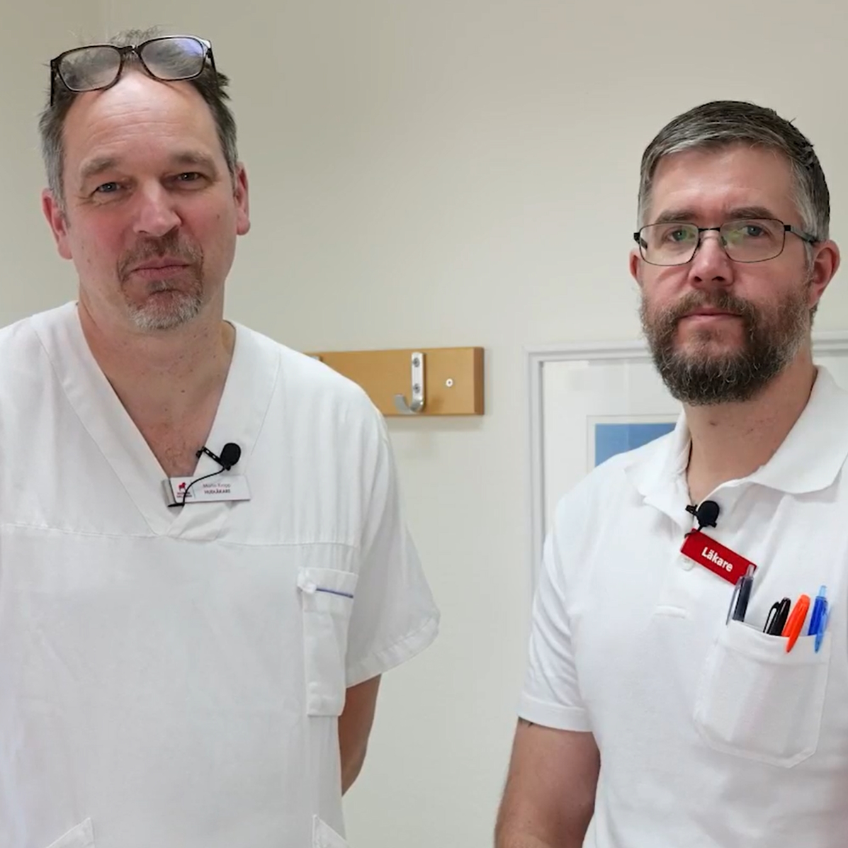
Can image review in VidiView lead to faster responses and a smoother care chain?
In Dalarna, they have managed to achieve a response time of within two days for most cases, reducing the burden on specialist care. The approach has been recognized, and in 2022, they received a regional award for General Practitioner Friend of the Year.
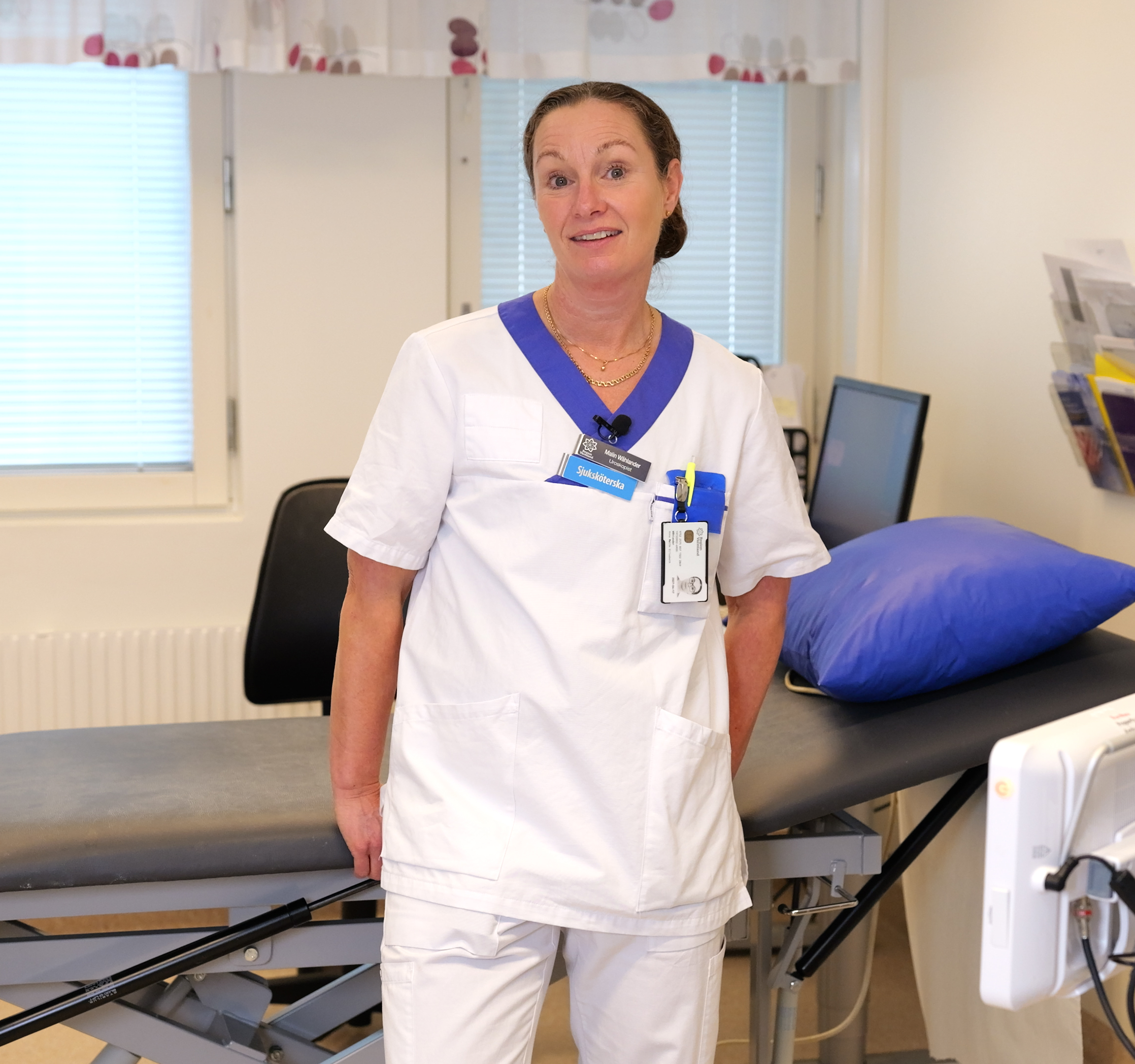
Can medical image documentation streamline workflows and enhance patient safety?
We had the opportunity to speak with Malin Wåhlander, a specialist nurse at the Cystoscopy Unit at Central Hospital in Karlstad. With extensive experience in the urology unit since 1997, Malin shares insights in our interview about how technological advancements and the implementation of VidiView have made workflows more efficient and improved patient safety.
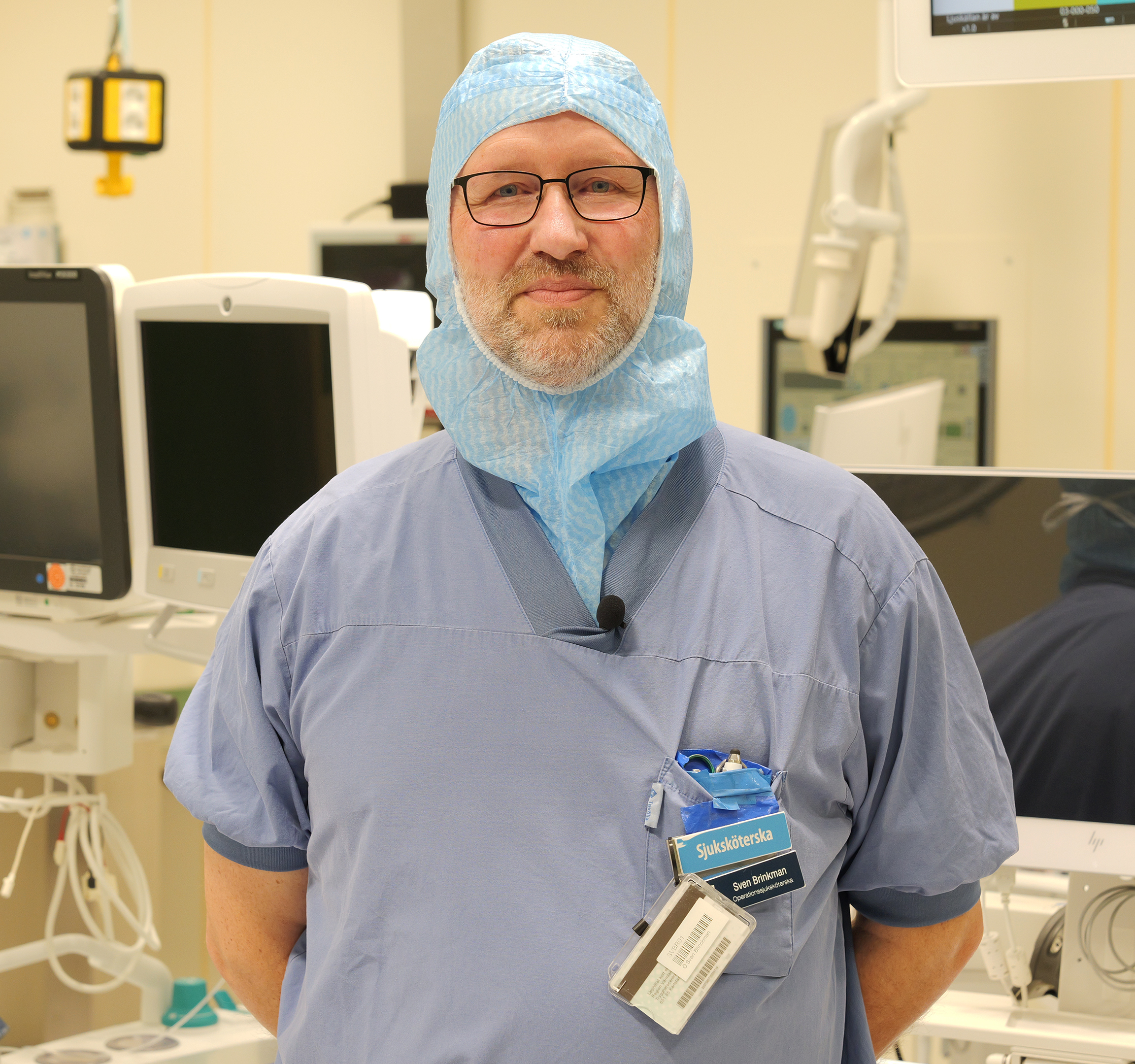
Can VidiView contribute to a better workflow in operating rooms?
We met with Sven Brinckmann, nurse and medical technology coordinator at the surgical department in Karlstad. Sven has worked with VidiView for six years and remembers how time-consuming it was to document surgeries in the past. Back then, the image material was saved on DVDs and USB sticks, which resulted in low image quality and inefficient handling.
Working with images – from primary care to hospital clinics
VidiView is widely used in Region Halland, everything from hospitals clinics to primary care units and even the private healthcare providers use the system today. The system is used by more than 5,000 users in the region.
“VidiView builds bridges between primary care centers and specialist care and helps shorten care queues”, she explains. “In Region Halland, we have chosen to include pictures in their referrals, e.g. when sending a referral from a primary care center to the dermatology clinic at the hospital. Then it becomes easier for the specialists to make decisions about referrals, which reduces incorrectly referred patients – who would otherwise have burdened the healthcare service unnecessarily. A true win-win!”
When implemented as broadly as in Region Halland – VidiView can truly help create a more efficient and safer workflow!
Video documentation as a patient safety toolbox
During a sit down with Lars-Göran Larsson, senior specialist consultant and surgeon at Kirurgkliniken in Mora (Sweden) we learned about his goal and vision with medical video documentation. In this video Lars Göran shares insight and experience form his long and extensive use of medical video documentation from the OR.
A well implemented video management system – the VidiView system in this case – can serve as a safety net for the surgeon. Helping him or her to resolve and discover problems, errors or mistakes from a surgery with a unforeseen or bad outcome.
The ability to, in a quick yet safe and secure way, bring up the video recording from the surgery greatly helps the investigation into what could have been done differently or better. The video is 100% objective and helps everyone involved remember exactly how the procedure was carried out in an unbiased way.
Lars-Görans method and vision is inspired by the aviation industry and their design with a ‘black box’ in the airplane – recording all events in case of a failure. A great help when sorting disasters. In both cases!
A well implemented video management system hence has at least 2 important roles to play – documentation and patient safety. At least if you ask Lars-Göran Larsson in Mora!
The history of surgical film making
We had the opportunity to sit down and talk to Lars-Göran Larsson, senior specialist consultant and surgeon at Kirurgkliniken in Mora (Sweden) about the history of medical image documentation and how medical images early on was used to leverage and educate surgical practice.
Lars-Göran is an avid medical documenter and is one of the main contributors to Videoarkivet USÖ. He has vast experience of medical video documentation and has worked with many different technical solutions in his mission. Today he prefers to work with the VidiView Enterprise Imaging solution deployed in the surgical clinic in Mora.
The recordings are done to enable review and develop the minimal invasive surgery methodology with the goal of increasing patient safety and reducing the number of patient injuries. The concept Lars-Göran develops is referred to as a Black box in the operating room (from the well-known method used in flight disaster recovery work).
With a simple push of a button, Lars-Göran and his colleagues can record and watch the surgery to analyze execution and methodology, train new surgeons and, from time to time, show the patient what it looked like during the surgery. Something that often is much appreciated!


